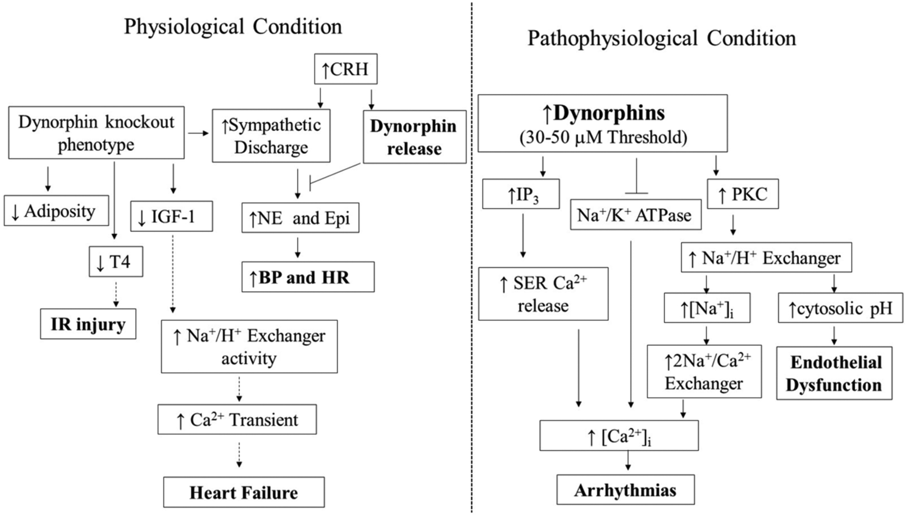
Fig. (2).

Schematic interpretation of the importance of dynorphins under physiological and pathophysiological processes in the cardiovascular system. Dynorphin fragments (Dyn A 1–17, Dyn A 1–13, and Dyn B 1–13) are involved in the regulation of tissues within the CV system. Panel A. Dynorphin A is cardioprotective. Dyn A 1–17 and Dyn A 1–13 dampen the sympathetic-induced increases in blood pressure and heart rate. Dyn deficient mice exhibit increased sympathetic activation. Lack of Dyn causes reduced IGF-1, T4, and free fatty acid levels. IGF-1 is capable of inhibiting Na+/H+ antiporter. Thus, increased IGF-1 may increase the development of heart failure via uncontrolled Na+/H+ antiporter hyperactivity-mediated increased calcium transient. Increased T4 protects cardiac tissue following I/R injury. Panel B. Arrhythmogenic signaling of dynorphins. Once a threshold is reached (30–50 μM), dynorphins induce arrhythmias through PKC- and IP3 - dependent mechanisms. Lastly, inhibition of Na+/K+ ATPase by Dyn leads to arrhythmias via intracellular calcium mobilization and activation of PKC -dependent Ca2+ signaling pathway. The flow of physiological changes that require further investigations are shown in dashed arrows. BP; blood pressure, CRH; corticotropic-releasing hormone, Epi; epinephrine, FFAs; free fatty acids, HR; heart rate, IGF-1; insulin-like growth factor 1, IP3; NE; norepinephrine, PKC; protein kinase C, SER; sarco/endoplasmic reticulum-associated calcium storage organelle, T4; thyroxine.
