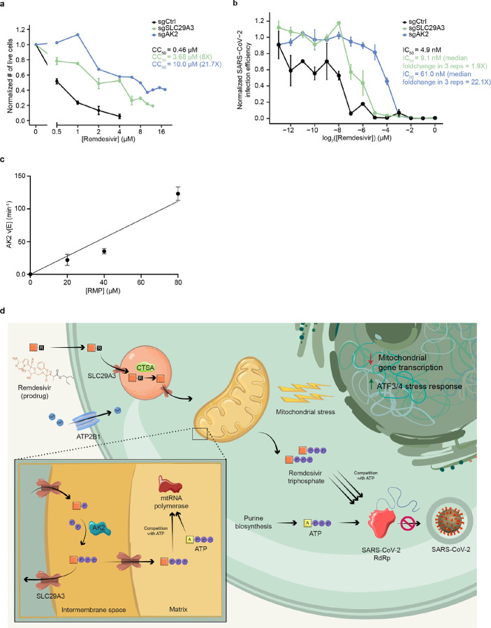
Figure 3. SLC29A3 knockout mitigates remdesivir toxicity without commensurate loss of SARS-CoV-2 antiviral potency, and AK2 is a remdesivir kinase required for antiviral potency and cytotoxicity.
a) Effect of remdesivir on average normalized number of live cells and CC50 in Huh7 sgCtrl and KO cell lines. b) Effect of remdesivir on average normalized SARS-CoV-2 infection efficiency and IC50 in sgCtrl and KO cell lines. c) Steady-state kinetic plot for AK2 catalytic rate (v/[E]) versus [RMP] (0–80 μ M). Reactions were carried out with 40 nM AK2 at 30° C for 10 minutes. d) Schematic of genes and pathways involved in the cellular metabolism and toxicity of remdesivir. The remdesivir prodrug is metabolized sequentially by Cathepsin A in the lysosome and AK2 in the mitochondrial intermembrane space into its active remdesivir-triphosphate form, which restricts SARS-CoV-2 RdRp in competition with ATP. SLC29A3 may modulate remdesivir-triphosphate entry into the mitochondrial matrix where its inhibition of mitochondrial RNA polymerase leads to toxicity. Remdesivir treatment leads to repressed nuclear transcription of mitochondrial genes, especially those involved in respiration, as well as leading to an ATF3/4 stress response. The plasma membrane transporter ATP2B1 is also involved in remdesivir cytotoxicity through an unknown mechanism.

