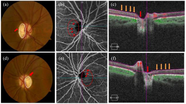
Figure 3.
Shows images of the right and left eyes in a patient with Wolfram syndrome (WS). Fundus photographs (a, d) show temporal pallor (white circle) and disc edema (red arrow) in the right (a) and left eyes (d). OCTA images of the superficial capillary plexus (b, e) demonstrate visible attenuation of the temporal microvascular networks of the optic nerve head (ONH) and peripapillary area (red circle) along with peripapillary telangiectatic blood vessels and vascular tortuosity (red arrows) for the right (b) and left eyes (e). OCT cross-sections (c, f) overlaying retinal flow (red) on OCT reflectance (gray scale) show a perfusion defect associated with ONH (red arrow) and temporal peripapillary nerve fiber layer (orange arrows) for the right (c) and left (f) eyes from Asanad and colleagues.98

