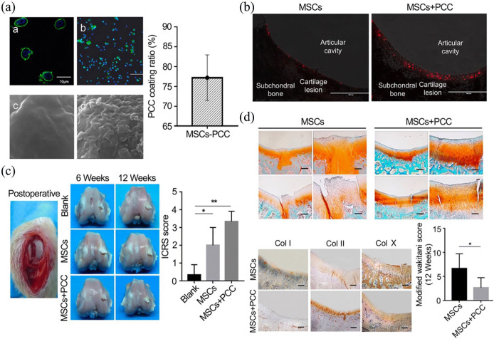
Figure 3.
Col I coating on BM-MSCs after intra-articular injection for cartilage defect. (A) Pericellular Col I coating. (a) Laser confocal microscopy observation of BM-MSCs coated with PCC. Scale bar = 10 μm. (b) Observation of PCC-coated BM-MSCs by fluorescence microscope. Scale bar = 500 μm. (c) SEM image of BM-MSC at 10,000× magnification. (d) SEM image of Col I–coated BM-MSCs at 10,000× magnification. (B) Homing and retention of BM-MSCs in vivo. Red particles represented BM-MSCs. (C) Macroscopic observation and histological scoring of the repaired cartilage. (D) Safranin O/fast green staining of regenerated cartilage and the modified Wakitani score of the regeneration of cartilage and immunohistochemical staining of Col I, Col II, and Col X in cartilage defect of trochlear groove after intra-articular injection of BM-MSCs and BM-MSC-PCC for 12 weeks. Scale bar = 100 μm. Adopted from Xia et al.66 and reprinted with permission of Springer Nature.

