Figure 1.
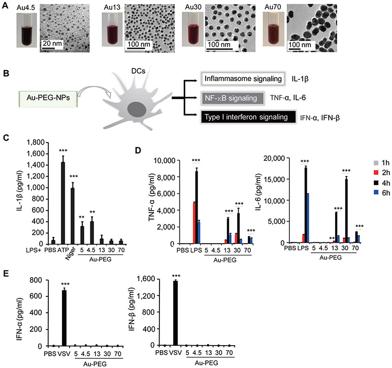
Size-dependent innate immune responses are induced by nanoparticles. (A) Optical images of Au nanoparticle suspension and transmission electron microscopy analysis of Au nanoparticles (top row). Scale bar presents as indicated. (B) Scheme for the screening strategy for innate immune responses to nanoparticles. (C) ELISA for the induction of IL-1β in supernatants of bone marrow derived dendritic cells (BMDCs) primed with LPS (100 ng/mL, 3 h) alone or followed by Au-PEG-NPs treatment at 200 μg/mL for 6 h. ATP was added at 5 mM for 1 h. Nigericin was added at 1 μM for 4–6 h. Mean diameter of each nanoparticle is indicated. (D) ELISA test for secretion of TNF-α and IL-6 in the supernatants of BMDCs stimulated with indicated Au nanoparticles (200 μ/mL) or LPS (100 ng/mL) for 1, 2, 4, and 6 h. (E) ELISA test for induction of IFN-α and IFN-β in supernatants of BMDCs stimulated with Au nanoparticles (200 μg/mL) or vesicular stomatitis virus (VSV) for 24 h. *p < 0.05, **p < 0.01, ***p < 0.001; NS, not significant. Data are representative of three experiments (mean ± SD).

