Figure 3.
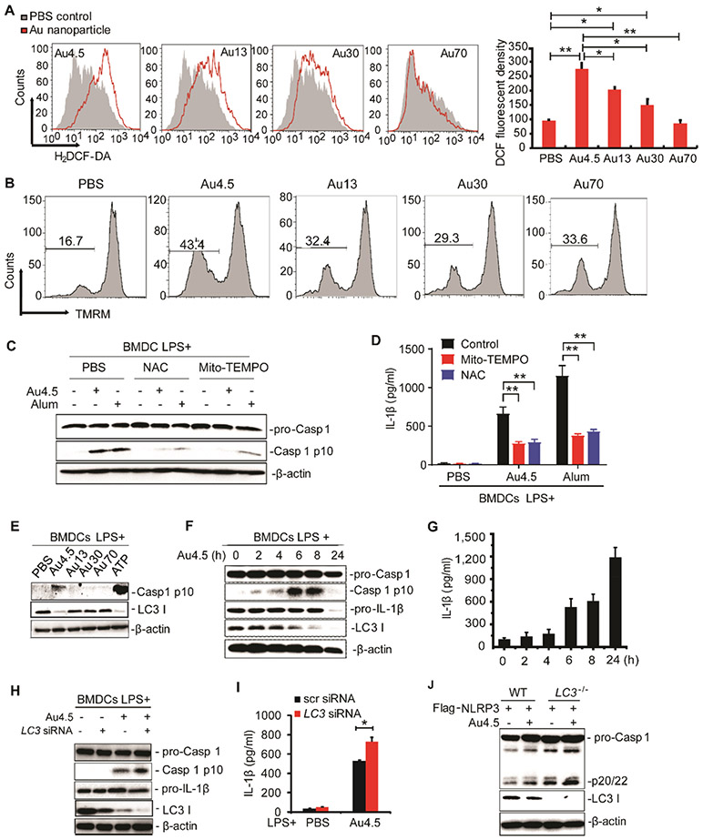
Au4.5 nanoparticles activate the NLRP3 inflammasome through ROS production and targeting LC3 for degradation. (A) Flow cytometry analysis of BMDCs’ intracellular ROS. BMDCs were labeled with H2DCF-DA to trace the intracellular ROS levels after incubation with 200 μg/mL of Au4.5, 13, 30, and 70 nanoparticles for 6 h, respectively. Quantities of fluorescent density are normalized from two independent experiments. (B) Flow cytometry analysis of BMDCs’ mitochondria depolarization using fluorescent probe TMRM upon treatment with 200 μ/mL of Au4.5, 13, 30, and 70 nanoparticles for 6 h, respectively. (C,D) Immunoblot analysis of cleaved caspase 1 in cell lysates (C) and ELISA for IL-1β in supernatants (D) of LPS-primed BMDCs, which were pretreated with MitoTEMPO (1 μM) or NAC (2 mM) for 3 h, followed by incubation with 200 μg/mL of Au4.5 or 400 μg/mL of alum for 6 h along with the MitoTEMPO or NAC. (E) Immunoblot analysis of cell lysates of BMDCs primed with LPS (100 ng/mL, 3 h) and then left untreated or followed by 6 h of Au4.5, Au13, Au30, or Au70 (200 μg/mL) treatment or 1 h of ATP treatment. (F,G) Immunoblot analysis of cell lysates (F) and ELISA for IL-1β in supernatants (G) of BMDCs primed with LPS (100 ng/mL, 3 h), followed by treatment with 200 μg/mL of Au4.5 for a different time. (H,I) Immunoblot analysis of pro-Caspase-1, Caspase-1 p10, and LC3 in cell lysates (H) and ELISA for IL-1β production in cell supernatants (I) of BMDCs transfected with scramble siRNA or LC3 siRNA and then primed with LPS (100 ng/mL, 3 h) alone or followed by 6 h of Au4.5 (200 μg/mL) treatment. (J) Immunoblot analysis of pro-Caspase-1, Caspase-1 p20/22, and LC3 in lysates of wild-type (WT) 293CIA cells or LC3 knockout 293CIA cells treated with Au4.5 (200 μg/mL) for 6 h. Data are representative of three experiments (mean ± SD). *p < 0.05.

