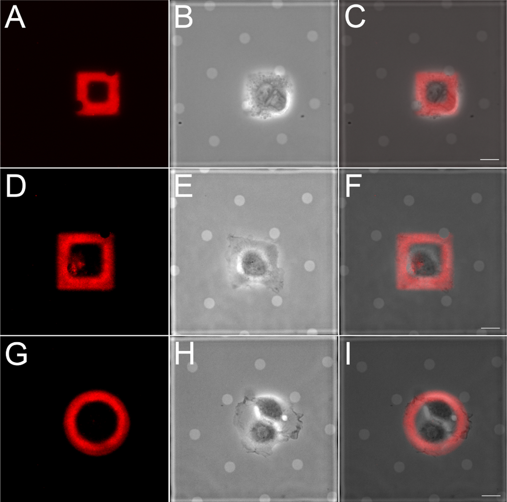
Figure 5:

Epithelial cells are confined to ECM patterns on EM grids generated by maskless photopatterning. PtK1 cells were plated on rhodamine-fibronectin square (A-C, 24.8 μm wide; D-F, 32.4 μm wide) and circle (36.6 μm wide) patterns, allowed to adhere and spread for 12 hr. (A, D, G) Rhodamine-fibronectin; (B, E, H) phase contrast; (C, F, I) overlay. Scale bar, 10μm.
