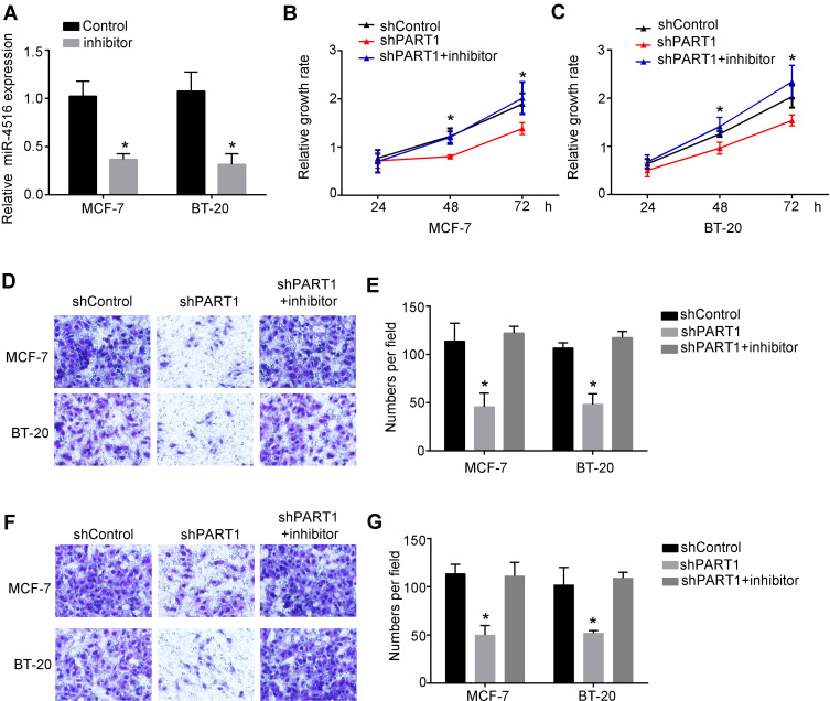
Figure 4.
PART1 promoted breast cancer cell progression through inhibiting miR-4516. (A) Relative expression of miR-4516 was detected by qRT-PCR after inhibition of miR-4516. Fold change was normalized to 18S. (B) Relative growth rate of MCF-7 cells and BT-20 cells were detected by Cell Counting Kit-8 (CCK-8) assay. Cells were transfected with shPART1 or shPART1 together with miR-4516 inhibitor or negative control shRNA. (C) Invasion abilities of MCF-7 cells and BT-20 cells were examined by transwell assay. MCF-7 cells and BT-20 cells were transfected with negative control shRNA, shPART1 or shPART1 together with miR-4516 inhibitor. (D and E) Migration abilities of MCF-7 cells and BT-20 cells were examined by transwell assay. MCF-7 cells and BT-20 cells were transfected with negative control shRNA, shPART1 or shPART1 together with miR-4516 inhibitor. (F and G) Invasion abilities of MCF-7 cells and BT-20 cells were examined by transwell assay. MCF-7 cells and BT-20 cells were transfected with negative control shRNA, shPART1 or shPART1 together with miR-4516 inhibitor. *P<0.05. All experiments were repeated three times.

