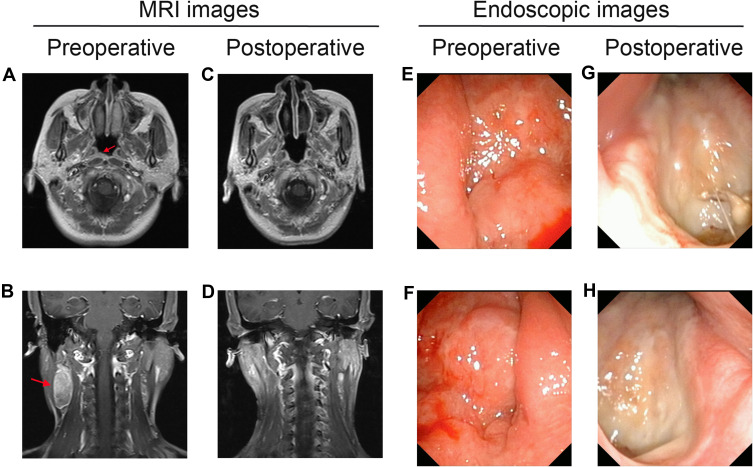
Figure 6.
Pre- and post-operative MRI and high-definition endoscopic images. (A) Pre-operative MRI shows that the tumor is located in the right pharyngeal recess (red arrow). (B) Pre-operative MRI shows that lymph node (LN) metastases is located in the right side of the neck (red arrow). (C, D) The post-operative MRI did not show tumor residual. (E) Pre-operative endoscopic examination shows the tumor is located in the left nasopharynx. (F) Pre-operative endoscopic examination shows the tumor is located in the right nasopharynx. (G, H) The post-operative endoscopic examination images show no visible tumor residual.

