. 2020 Jul 7;296(3):641–649. doi: 10.1148/radiol.2020192429

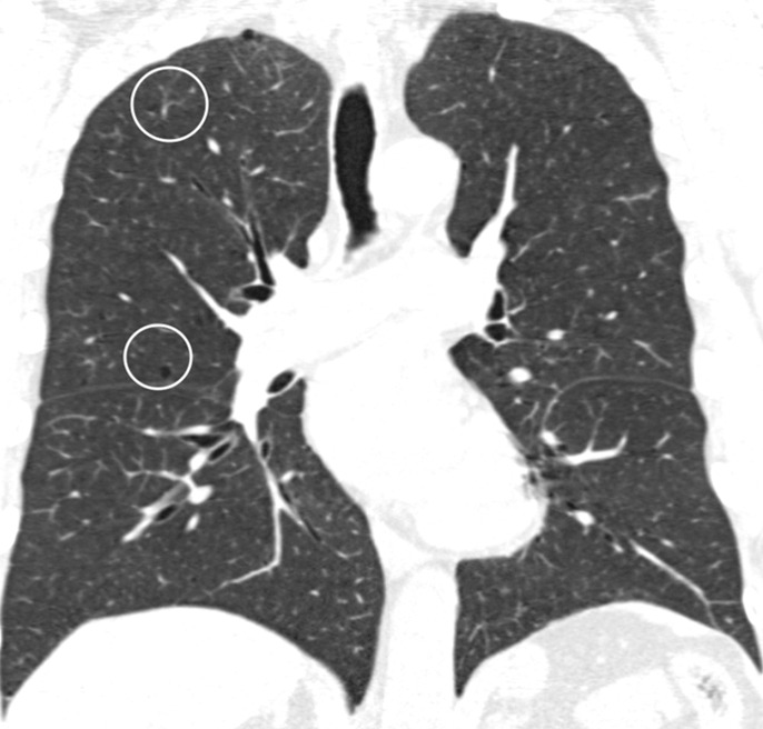
Figure 2b:

Coronal CT images show progressive grades of parenchymal emphysema on the basis of the Fleischner classification system. (a) Normal CT scan shows no emphysema. (b) Trace centrilobular emphysema (circles). (c) Mild centrilobular emphysema (arrows). (d) Moderate centrilobular emphysema involving more than 5% of the lung zone. (e) Confluent emphysema. (f) Advanced destructive emphysema.

Coronal CT images show progressive grades of parenchymal emphysema on the basis of the Fleischner classification system. (a) Normal CT scan shows no emphysema. (b) Trace centrilobular emphysema (circles). (c) Mild centrilobular emphysema (arrows). (d) Moderate centrilobular emphysema involving more than 5% of the lung zone. (e) Confluent emphysema. (f) Advanced destructive emphysema.