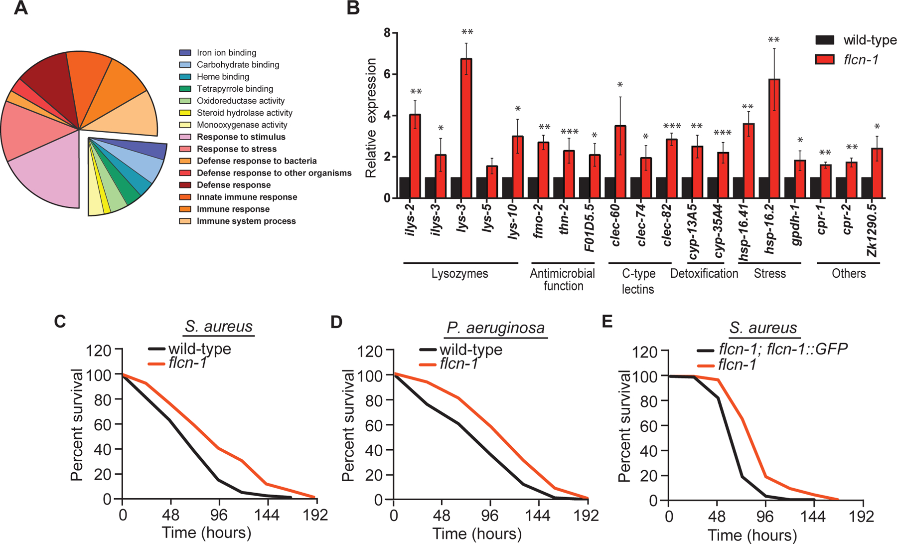
Figure 1: Loss of flcn-1 increases the expression of antimicrobial genes and confers resistance to bacterial pathogens.

(A) Pie chart of functional gene ontology analysis of the genes upregulated in flcn-1(ok975) mutant animals at basal level. (B) Relative mRNA expression of stress response and antimicrobial peptide genes in wild-type and flcn-1 mutant animals. Data represents the average of three independent experiments, each done in triplicates ± SEM. Significance was determined using student’s t-test (*p<0.05, **p<0.01, ***p<0.001). (C-E) Percent survival of indicated strains upon infection with S. aureus and P. aeruginosa. Refer to Table S1 for details on number of animals utilized and number of repeats. The Statistical analysis was obtained using Mantel Cox test on the pooled curve.
