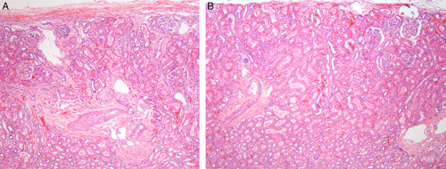
FIGURE 11.

A, This image in a case of segmental hypoplasia from an 11-year-old shows cortical hypoplasia in a lobe adjacent to a hypoplastic focus. Only 2 nephron generations are present. Normal-appearing outer medulla is visible below the arteries in each image. B, This is a second image in a case of segmental hypoplasia from the same 11-year-old that showed cortical hypoplasia in a lobe adjacent to another hypoplastic focus. Only 2 nephron generations are present. Normal-appearing outer medulla is visible below the arteries in each image.
