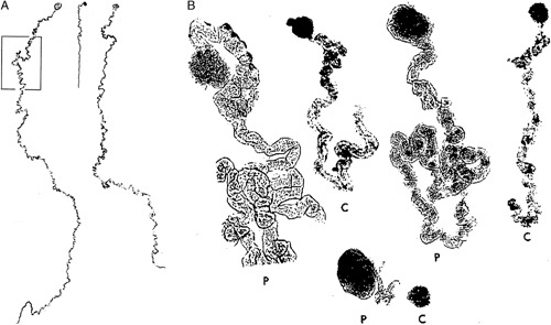
FIGURE 7.

A, This is a mosaic photograph of 2 glomeruli with proximal tubules microdissected from a kidney of a patient with oligomeganephronia, left and right side. These are compared with an age-matched control glomerulus with proximal tubule shown in the center. Reprinted from Fetterman and Habib,35 Fig. 2 with permission from Oxford University Press. Copyright Oxford University Press, New York, NY. All permission requests for this image should be made to the copyright holder. B, This image shows glomeruli and proximal tubules (P) microdissected from a patient with oligomeganephronia compare to an age-matched control (C). Reprinted from Griffel et al,47 Fig. 2 with permission from Springer Nature. Copyright Springer Nature, New York, NY. All permission requests for this image should be made to the copyright holder.
