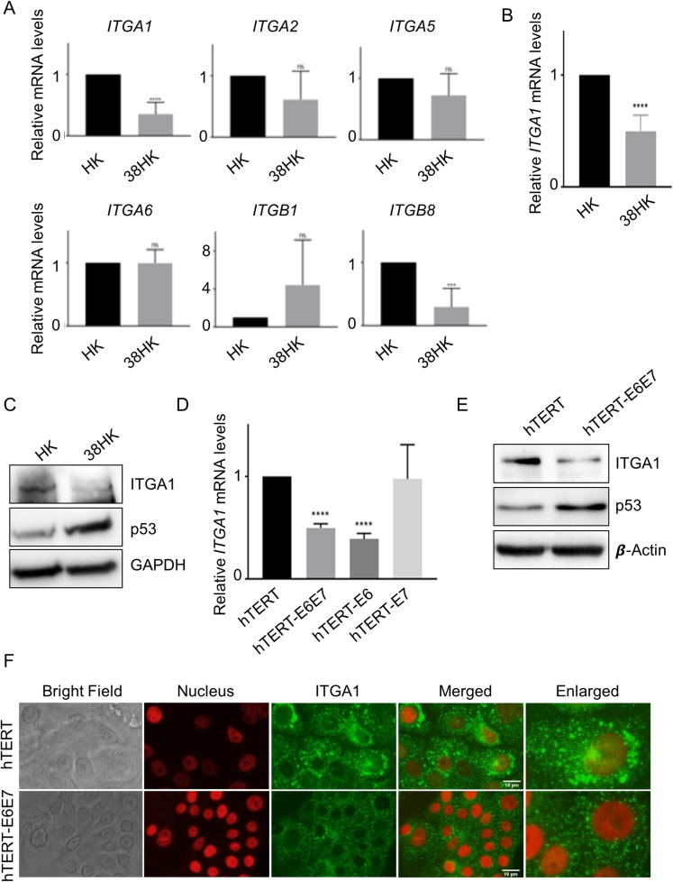
Fig 1. ITGA1 expression is downregulated in HPV38 E6/E7-expressing cells.
(A) Primary HKs were transduced with pLXSN HPV38 E6/E7 or pLSXN. mRNA levels were measured by RT-qPCR and normalized to GAPDH. Error bars represent standard deviations from 3 biological replicates of 2 different donors (n = 6). ***, p<0.001; ****, p<0.0001; ns, not significant. (B) Total RNA levels of HKs expressing or not expressing HPV38 E6 and E7 were analyzed by TaqMan PCR. Commercial probes for ITGA1 and GAPDH were used. Results were normalized to GAPDH. Data shown are the means of 3 independent experiments for 2 different donors (n = 6). ****, p<0.0001. (C) Proteins extracts from HKs expressing or not expressing HPV38 E6 and E7 were analyzed by immunoblotting (IB) with the indicated antibodies. (D) The TaqMan assay was also performed as previously described in primary HKs previously retrovirally transduced with the hTERT gene and expressing E6 and/or E7 from HPV38 (n = 3). Results were normalized to GAPDH. ****, p<0.0001. (E) Proteins extracts from hTERT pLXSN or hTERT HPV38 E6/E7 cells were analyzed by IB with the indicated antibodies. Images shown are representative examples of 2 different experiments. (F) hTERT pLXSN or hTERT HPV38 E6/E7 cells were plated on coverslips and after 24 h were probed for ITGA1 using anti-ITGA1 antibody followed by secondary Alexa Fluor 488-conjugated antibody. Nuclei were stained with DAPI (pseudocoloured red), and cells were analyzed under a microscope. Images were merged using ImageJ software.

