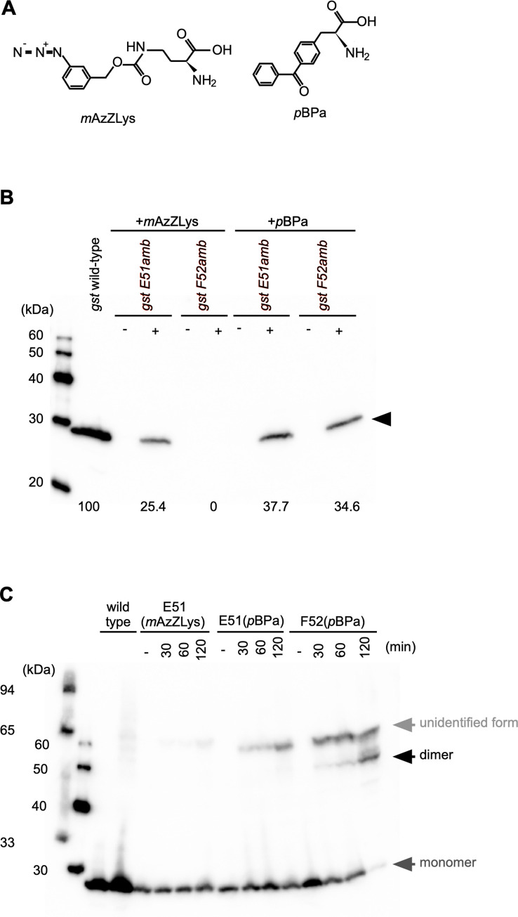
Fig 3. Incorporation of ncAAs into N. meningitidis proteins monitored by Western blotting.
(A) Chemical structures of ncAAs, mAzZLys (Left) or pBPa (right) used in this study. (B) Estimation of the efficiency of pyrrolysine-based amber suppression with mAzZLys or pBPa for GST E51amb or GST F52amb in N. meningitidis. Bacterial extracts equivalent to an OD600 of 0.1 were analyzed by Western blotting with an anti-GST mAb. The horizontal black arrow shows the full-length GST protein. Numbers indicate relative suppression efficiency when wild-type GST expression is defined as 100%. (C) Detection of irradiation- and time-dependent GST dimerization of GST F52 (pBPa) in N. meningitidis. Bacterial extracts equivalent to an OD600 of 0.05, irradiated with UV light for the indicated times, were analyzed by Western blotting with an anti-GST mAb. Dark gray and black arrows show the monomer and dimer forms of the GST protein, respectively. The light gray arrow shows an unspecified form of GST generated by UV irradiation.

