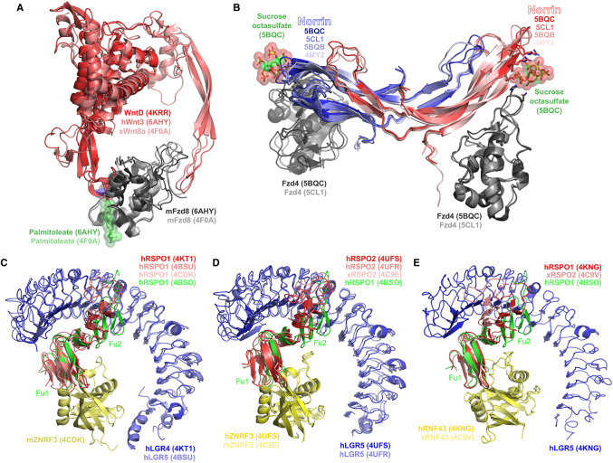
Figure 2. Structures related to interactions involving secreted positive regulators of Wnt signalling.
(A) Wnt-related proteins and interactions. Structures depicted include: WntD (PDB 4KRR); the human Wnt3 complex with the mouse Fzd8 cysteine-rich domain (PDB 6AHY); the Xenopus Wnt8a complex with the mouse Fzd8 cysteine-rich domain (PDB 4F0A). (B) Norrin and its interactions. Structures depicted include: unbound norrin-maltose binding protein fusion (PDB 4MY2), unbound norrin (PDB 5BQB), norrin-maltose binding protein fusion in complex with Fzd4 (PDB 5CL1), norrin-Fzd4-sucrose octasulfate ternary complex (PDB 5BQC). Maltose binding protein hidden. Key residues contacting sucrose octasulfate in PDB 5BQC are shown as sticks. (C) RSPO1–LGR–ZNRF3 complexes. Structure represented include: human RSPO1–LGR4 complex (PDB 4KT1), human RSPO1–LGR5 complex (PDB 4BSU), human RSPO1 in complex with mouse ZNRF3 (PDB 4CDK). The structure of native human RSPO1 (PDB 4BSO) is shown for reference. (D) RSPO2–LGR–ZNRF3 complexes. Structures represented include: human RSPO2–LGR5–ZNRF3 complex (PDB 4UFS), human RSPO2–LGR5 complex (PDB 4UFR), Xenopus RSPO2 in complex with mouse ZNRF3 (PDB 4C9E). The structure of native human RSPO1 (PDB 4BSO) is shown for reference. (E) R-spondin–LGR–RNF43 complexes. Structures represented include: human LGR5–RSPO1–RNF43 complex (PDB 4KNG); Xenopus RSPO2–RNF43 complex (PDB 4C9V). The native human RSPO1 (PDB 4BSO) is shown for reference.

