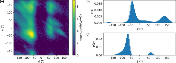
Figure 1. Illustration of projection of a free energy landscape onto commonly used CVs.
(a) Ramachandran maps project the conformational free energy landscape onto the backbone φ and ψ dihedral angle values. The example shown here is for a 100 ns MD simulation of hen egg white lysozyme (PDB ID: 1aki). (b,c) Projections of the conformational free energy landscape onto a single CV: (b) ψ and (c) φ. All angle values are in degrees. Projection of the free energy landscape onto the combination of both backbone dihedral angles is useful because it clearly separates the two major regions of secondary structure, namely (right-handed) α-helices and β-strands, although it is less effective at providing a more detailed degree of separation, such as between parallel and antiparallel β-strands — for this, additional CVs are required. ψ alone (b) could be a useful CV, as it preserves this separation, whereas projection onto φ (c) conflates α-helical and β-strand structure.

