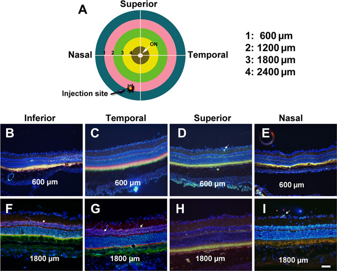
Fig. 2. Widespread expression of GFP in retina and RPE 2 months after suprachoroidal injection of NPs containing a CMV-GFP expression plasmid.

Brown Norway rats were given a 3-μl suprachoroidal injection of PBAE NPs containing 1 μg of CMV-GFP expression plasmid 1 mm posterior to the limbus in the inferonasal quadrant near the vertical meridian (A). Two months after injection, rats were euthanized, eyes were frozen in embedding medium, and transverse sections were immunohistochemically stained for GFP, counterstained with Hoechst, and merged with its fluorescent image. In the inferior (B), temporal (C), superior (D), and nasal (E) meridians of a section 600 μm posterior to the limbus through anterior retina [intersections of inner surface of blue ring and white lines in (A)], there was colocalization of fluorescence with anti-GFP staining in RPE and photoreceptor inner and outer segments. At the inferior (F), temporal (G), superior (H), and nasal (I) meridians of a section 1800 μm posterior to the limbus through posterior retina, staining for GFP in RPE and photoreceptor inner and outer segments was similar to that seen in anterior retina. Occasional weak expression of GFP was seen in cells of the inner retina (arrows). Scale bar, 100 μm.
