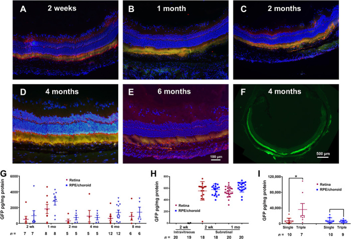
Fig. 3. Time course of GFP expression after suprachoroidal injection of NPs containing a CMV-GFP expression plasmid.

Brown Norway rats were given suprachoroidal, intravitreous, or subretinal injection of PBAE NPs containing 1 μg of CMV-GFP expression plasmid. Ocular sections immunohistochemically stained for GFP (Hoechst counterstain) merged with fluorescent image showed GFP in photoreceptor inner and outer segments and RPE 2 weeks (A), 1 month (B), 2 months (C), 4 months (D), and 6 months (E) after vector injection. Low magnification showed GFP fluorescence around the entire circumference of the eye (F). ELISA showed high levels of GFP in retina and RPE/choroid from 2 weeks to 8 months (last time point measured). (G). GFP was undetectable in retina and RPE/choroid 2 weeks after intravitreous injection of PBAE NPs, but there were high levels 2 weeks and 1 month after subretinal injection (H). Compared with eyes given a single suprachoroidal injection of PBAE NPs containing 1 μg of CMV-GFP, those given three injections had significantly higher levels of GFP in retina, but not RPE/choroid (I). *P = 0.018 by two-sample t test with equal variance for comparison with corresponding single injection control.
