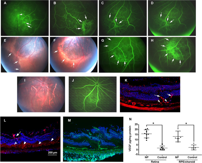
Fig. 4. Nonviral suprachoroidal gene transfer of Vegf causes subretinal NV progressing to subretinal fibrosis.
Rats were given suprachoroidal injection of PBAE NPs containing 1 μg of pVEGF. After 4 weeks, FA showed subretinal hyperfluorescence (A and B) (arrows) and leakage from retinal vessels (arrowhead). After 8 weeks, subretinal hyperfluorescence (C) (arrows) spread over a minute (D) (arrows), indicating leakage from subretinal NV. After 5 months, there was subretinal fibrosis (E and F) that stained with fluorescein (G and H) (arrows). An eye injected with control vector showed normal fundus (I) and FA (J). Four-month-old rho/VEGF mouse (K) showed new vessels from deep capillaries (arrows) to subretinal space (arrowheads; DyLight 594–conjugated GSA). Four months after suprachoroidal injection of NPs containing 1 μg of pVEGF, new vessels extended from deep capillaries to severe subretinal NV (L) and, in another eye, showed severe NV with disrupted retina (M) (fluorescein isothiocyanate GSA). Two weeks after suprachoroidal injection of 1 μg of pVEGF in NP, ELISA showed significantly higher VEGF in retina or RPE/choroid versus controls (N) (n = 8 NPs and n = 6 controls). *P < 0.0001 for retina and for RPE/choroid by two-sample t test with unequal variance for comparison with control. Scale bar, 200 μm.

