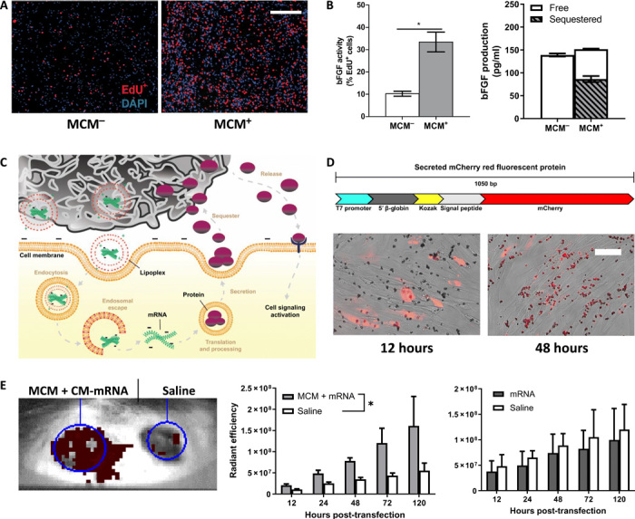
Fig. 2. MCMs extend biological effects of mRNA delivery via an “overexpress and sequester” mechanism.

(A) Representative epifluorescence micrographs of hDFs transfected with 150-ng bFGF CM-mRNA with (+) and without (−) MCMs, 48 hours after transfection. Scale bar, 500 μm. (B) Left: S phase+ cells (red) quantified as the percentage of total 4′,6-diamidino-2-phenylindole (DAPI; blue) nuclei. n = 3; *P ≤ 0.05, one-way Student’s t test. Right: bFGF production and sequestering after bFGF CM-mRNA transfection. “Free” represents bFGF in the media, and “sequestered” is the measured bFGF after mineral coating dissolution via EDTA. n = 3. (C) Schematic of overexpress and sequester mechanism. MCMs deliver mRNA lipoplexes and then bind and sequester the secreted protein, sustaining growth factor release over time and prolonging the biological response. (D) Schematic (top) for model secreted mCherry protein and (bottom) merged phase and epifluorescence micrographs of transfected hDFs 12 (left) and 48 (right) hours after transfection. Scale bar, 50 μm. (E) Representative IVIS image of the secreted mCherry CM-mRNA + MCM–treated dermal wound (left wound) and saline control (right wound) 48 hours after delivery. (F) Red fluorescence radiant efficiency within each transfected or saline wound site over five with MCMs (left) and without (right). n = 3; *P < 0.05 by two-way ANOVA.
