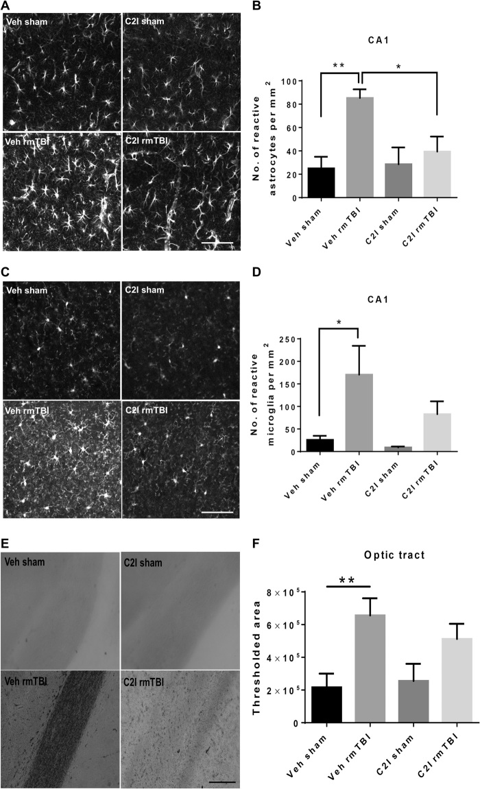
Fig. 5. Effects of C2I treatment on glial activation and axonal degeneration following repeated concussions in WT mice.
WT mice were implanted with Alzet minipumps delivering vehicle [veh; 400 mg/ml; (2-hydroxypropyl)-β-cyclodextrin] or C2I (0.3 mg kg−1 day−1) 1 day before 10 days of repeated concussions. Pumps were withdrawn 4 days after the last day of concussion, and the animals were sacrificed 4 weeks later. (A) Changes in astrocyte activation in field CA1 of hippocampus. Brains were fixed and processed for IHC with GFAP antibodies. Scale bar, 100 μm. (B) Quantification of images similar to those shown. n = 8 for veh sham and veh rmTBI, n = 7 for C2I sham, n = 9 for C2I rmTBI. **P < 0.01. One-way ANOVA followed by Bonferroni’s test. Data represent means ± SEM. (C) Changes in microglia activation in field CA1 of hippocampus. Brains were fixed and processed for IHC with iba-1 antibodies. Scale bar, 100 μm. (D) Quantification of images similar to those shown. n = 8 for veh sham and veh rmTBI; n = 7 for C2I sham; and n = 9 for C2I rmTBI. *P < 0.05. One-way ANOVA followed by Bonferroni’s test. Data represent means ± SEM. (E) Changes in axonal degeneration in the optic tract. Brains were fixed and processed for Gallyas staining. Scale bar, 100 μm. (F) Quantification of images similar to those shown. n = 6. **P < 0.01. One-way ANOVA followed by Bonferroni’s test. Data represent means ± SEM.

