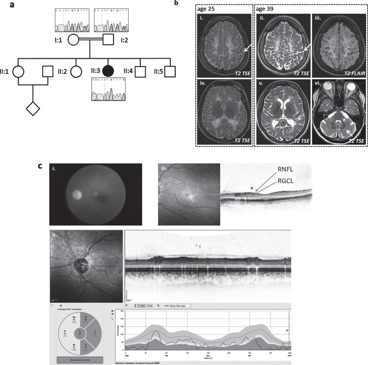
Fig. 2.
Patient 2, homozygous for c.252_253delCT (p.Cys85PhefsTer15). (a) Pedigree of family and Sanger sequencing show that the prob and is homozygous and her mother and father are heterozygous for the TACO1c.252_253delCT variant. (b) Baseline MRI image at the age of 25 years (i + iv on the left) and follow-up 14 years later (ii/iii+v/vi) show white matter degeneration with white matter atrophy and sulcal widening (arrows in i and ii) as well as cystic lesions that demonstrate CSF like signal behavior in the FLAIR (iii). The infratentorial T2-weighted image (vi)demonstrates hyperintensities within the middle cerebellar peduncle (asterix) and within the cerbellar white matter (arrow). (c) Color fundus photography (i) shows severe paleness of the optic nerve head. OCT (ii+iii) reveals atrophy of the peripapillary retinal nerve fiber layer (RNFL) (ii). The foveal depression is flattened due to atrophy of the ganglion cell layer (GCL) and retinal nerve fiber layer (RNFL) (see * in (iii)). All other retinal layers are intact; in particular, there are no signs or chorioretinal atrophy. Best corrected visual acuity was 20/400 (right eye) and 20/250 (left eye), the subject was legally blind (according to WHO definition). Images for the left eye are shown; all changes were present in both eyes.

