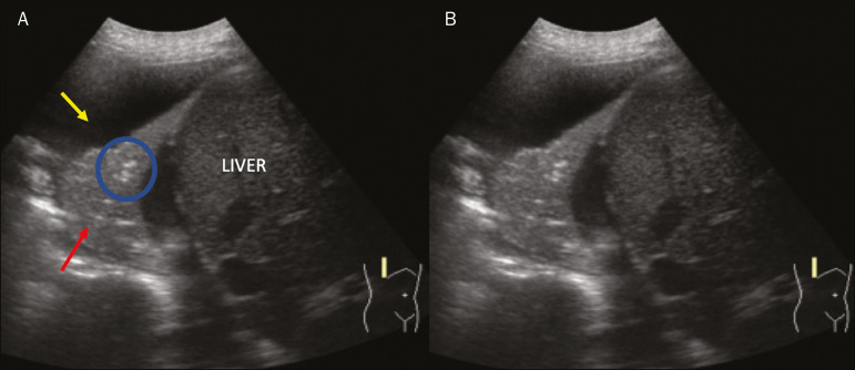
Figure 10.
A,B: Ultrasound view of the lower third of the right hemithorax. Atelectasis (red arrow) and pleural effusion (yellow arrow). Note the volumetric reduction of the pulmonary segment assessed and the presence of static air bronchogram (circle). During respiratory incursions, there is no change in its characteristics and in the Doppler phenomenon, which would be present in the dynamic bronchograms of consolidations.

