
Figure 4.
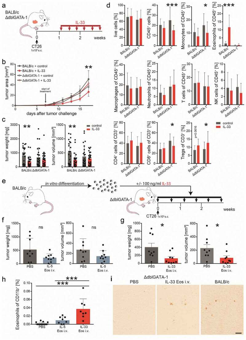
Eosinophils are necessary for a reduction in tumor growth by IL-33.
(a) Schematic presentation of the subcutaneous (s.c.) tumor model performed in BALB/c and eosinophil deficient ∆dblGATA-1 mice. IL-33 was given i.p. at a concentration of 0.4 µg/mouse every other day for a total of six times. (b) Tumor growth was monitored during the course of the experiment; n ≥ 8. Data indicate mean values ± SEM. **p < .01 BALB/c + control vs. BALB/c + IL-33; no significance between other groups. (c) One day after the last IL-33 injection, BALB/c and ∆dblGATA-1 mice were sacrificed and tumor weight and volume were measured ex vivo showing no differences between IL-33- and vehicle (control) treatment in the ∆dblGATA-1 but tumor growth reduction in the BALB/c mice; n ≥ 23, three independent experiments. (d) Flow cytometric analysis of single cell suspensions of tumors from BALB/c and ∆dblGATA-1 mice indicating significant differences between IL-33- and vehicle-treated (control) ∆dblGATA-1 mice for CD45+, monocytes, CD8+ T cells and Tregs, as well for eosinophils between IL-33- and vehicle-treated (control) BALB/c mice; n = 24; three independent experiments. (e) ∆dblGATA-1 mice with s.c. tumors were repopulated by adoptive transfer (twice weekly) with either IL-33 Eos or IL-5 Eos (eosinophils were isolated from bone marrow of BALB/c mice and differentiated). (f) Adoptive transfer with IL-5 Eos failed to significantly restore tumor reduction in ∆dblGATA-1 mice (in comparison to ∆dblGATA-1 mice given PBS only). (g) However, adoptive transfer with IL-33 Eos (vs. PBS only) significantly restored reduction of tumor growth; n = 8–9. (h) Single cell suspensions of the s.c. tumors were prepared and infiltrated eosinophils were evaluated as % of CD11b+ cells; n = 8–15. (i) Immunohistochemistry of s.c. tumors with an anti-EPX antibody shows that PBS-treated ∆dblGATA-1 mice are devoid of eosinophils. After adoptive transfer of IL-33 Eos (by i.v. injection), EPX staining is visible in the tumors of ∆dblGATA-1 mice. Infiltrated eosinophils in s.c. tumors of BALB/c mice are shown for comparison (representative images of n = 3/group; calibration bar: 20 µm). Statistical differences were assessed by using multiple t-tests, one-way ANOVA with Tukey’s multiple comparisons test and unpaired student’s t-test. *p < .05; **p < .01; ***p < .001, ns = not significant.