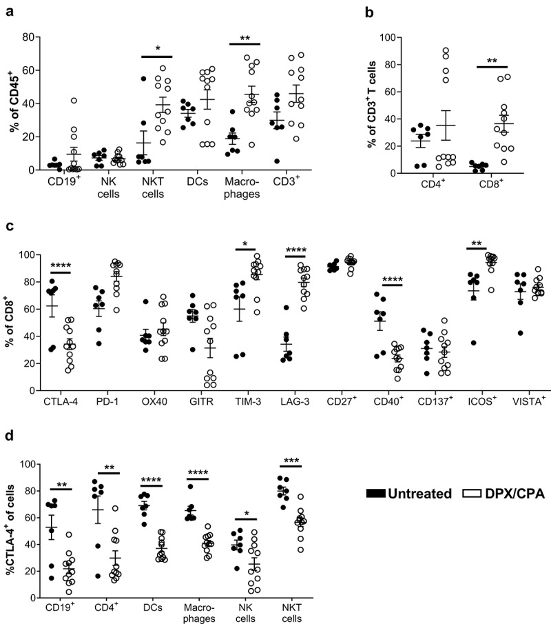
Figure 1.

'Flow cytometry analysis suggests DPX/CPA treatment causes a decrease in CTLA-4+ tumor-infiltrating cells into the C3 tumor.
Mice were implanted with tumors on day 0 and treated with CPA 14 days later for a week. DPX-FP was administered on day 21. Ten days post DPX-FP treatment (study day 31), mice were terminated. Tumors were dissociated and analyzed by flow cytometry to characterize different immune cell populations and their expression of checkpoint markers. a) B cells, NK cells, NKT cells, dendritic cells, macrophages, and T cells of CD45+ cells. Note each cell type was analyzed on separate panels; therefore, the total of each cell type does not add up to 100%; b) Percent CD4+ and CD8+` of total T cells; c) Percent of checkpoint marker positive of CD8+ T cells; d) Percent of CTLA-4+ of tumor-infiltrating immune cells. Results pooled from three separate experiments, n = 7–11, average ± SEM, statistics by student's t-test, * p<0.05, ** p< 0.01, *** p<0.001, **** p<0.0001
