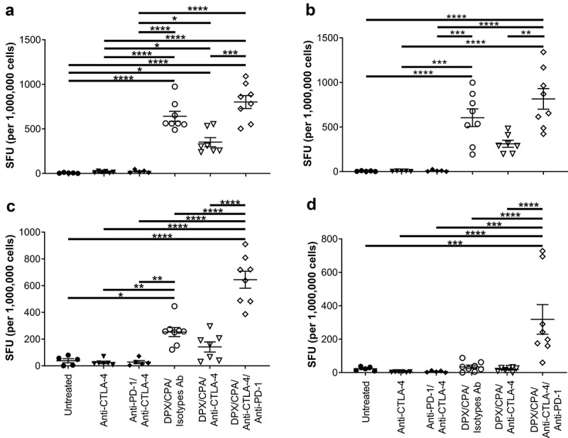
Figure 7.

CTLA-4 blockade does not enhance the impact DPX/CPA treatment has on antigen-specific IFN-γ production.
Experimental design same as stated in Figure 4. Spleens and vaccine-draining lymph nodes were collected for immune analysis by IFN-γ ELISPOT assay. Splenocytes (A, C) stimulated with R9F peptide (A), or C3 cells (C). Number of SFU in untreated splenocyte samples were ≤89. Lymph node (B, D) cells were stimulated with R9F peptide-loaded syngeneic dendritic cells (B), or C3 cells (D). Number of SFU in untreated lymph node samples in untreated and antibody-alone groups, and DPX/CPA/Isotype antibodies (Ab) or anti-CTLA-4 was ≤162.5; number of SFU in untreated lymph node samples in DPX/CPA/anti-CTLA-4/anti-PD-1 ranged from 55 to 640. Untreated, irrelevant R9L, and irrelevant Panc02 data presented in Supplementary Figure 5. Order of samples in all panels: untreated (filled circles); anti-CTLA-4 (filled down-pointing triangles); anti-PD-1/anti-CTLA-4 (filled diamonds); DPX/CPA/Isotypes Ab (empty circles); DPX/CPA/anti-CTLA-4 (empty down-pointing triangles); DPX/CPA/anti-CTLA-4/anti-PD-1 (empty diamonds). n = 5–8, average ± SEM, statistics by one-way ANOVA followed by a Tukey’s multiple comparisons posttest, * p<0.05, ** p<0.01, *** p<0.001, **** p<0.0001
