
Figure 3.
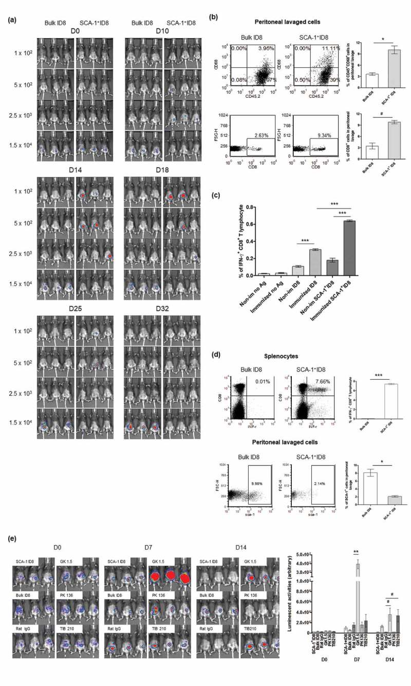
SCA-1+ ID8 cells were immunogenic and being rejected in vivo by anti-tumor immunity involving CD4+ and CD8+ T lymphocytes. (a) SCA-1+ ID8 cells showed stronger tumorigenesis initially by D18 but disappeared later. Bulk ID8 cells grew slower but did form tumor eventually by day 25. (b) Representative flow analysis of CD8+ and CD68+ cells in peritoneal lavage of mice injected with 2.5 × 10³ bulk ID8 and SCA-1+ ID8 cells on D22. The results show significantly increased percentages of CD45+CD68+ cells and CD8+ cells in peritoneal lavage from mice implanted with SCA-1+ ID8, comparing to mice with bulk ID8 cells. (c) Analysis of splenocytes demonstrated the highest number of IFN-γ+ CD8+ T lymphocytes was found in mice in which SCA-1+ tumors disappeared (splenocytes from mice injected with 2.5 × 10³ SCA-1+ cells on D32, as immunized) and were re-stimulated with irradiated SCA-1+ ID8 cells (p < .0001, splenocytes re-stimulated by irradiated bulk ID8 cells [5 × 10⁴ cell] [labeled as immunized ID8] vs splenocytes re-stimulated by irradiated SCA-1+ ID8 cells [5 × 10⁴ cells] [labeled as immunized SCA-1+ ID8]). Splenocytes from those mice with SCA-1+ tumor disappeared also showed higher percentages of IFN-γ+ CD8+ T lymphocytes than that from mice injected with 2.5 × 10³ cells bulk ID8 cells, irrespective of the re-stimulated antigen (p < .0001, immunized ID8 vs non-immunized ID8 and immunized SCA-1+ ID8 vs non-immunized SCA-1+ ID8). (d) Representative data show that mice immunized with 3 doses of irradiated SCA-1+ID8 cells exhibited strong tumor-specific CD8+ T lymphocyte response (p < .0001) and rejected the growth of SCA-1+ cells inside the peritoneum (p < .01). Rejection of SCA-1+ tumor was more obviously seen in mice immunized with irradiated SCA-1+ ID8 cells demonstrated by less SCA-1+ lavaged cells in peritoneal cavity. (e) Rejection of SCA-1+ ID8 tumor was abolished by CD4+ and CD8+ lymphocyte depletion. All C57BL/6 mice were implanted with SCA-1+ ID8 cells, except those labeled with “Bulk ID8” were implanted with bulk ID8 cells. Lymphocyte depletion was performed through i.p. injection of rat monoclonal antibody GK1.5 (anti-CD4), 2.43 (anti-CD8), or PK136 (anti-NK1.1) described in Materials and Methods. Signal of SCA-1+ tumor peaked on D7 if CD4+ lymphocyte was neutralized by GK 1.5 antibody. The signals were reduced on D14, possibly through the immune response elicited by rapidly growing tumor. SCA-1+ tumor became obvious on day 14 if CD8+ lymphocyte was neutralized by TIB 210 antibody. Administration of rat IgG or PK136 antibody did not alter the rejection of SCA-1+ tumors. These data suggest involvement of CD4+ and CD8+ immune reactions in the attack of SCA-1+ ID8 stem-like cells. #p < .05, *p < .01, **p < .001, ***p < .0001.