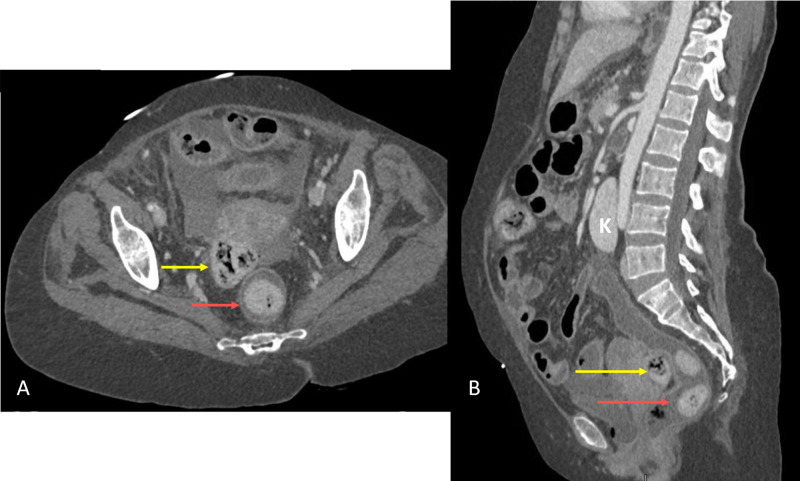
Figure 2. Contrast-enhanced CT scan of the abdomen, composite view.
(A) shows the axial view and (B) shows the sagittal view. Red arrows in (A) and (B) show intraluminal fecalomas in the rectum. Yellow arrows show extraluminal faecal ball in the peritoneal cavity in the Pouch of Douglas. Isthmus of the horseshoe kidney (K) was connecting the lower poles and is seen overlying the aorta

