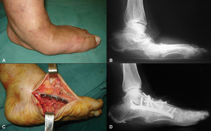
Fig. 1.

Clinical aspect ( A ) and radiographic image of a patient with Charcot neuroarthropathy , in lateral view of the left foot, with a support base ( B ), affecting the midfoot (type I of the anatomical classification of Brodsky 4 and Trepman 22 ). Note the pronounced abduction deformity ( A ) and severe collapse of the arch with plantar bony prominence (A and B). Surgical treatment consisted of modeling midfoot arthrodesis through medial approach for bone wedge resection and correction of deformities ( C ), followed by internal fixation with plate and screws ( D ).
