. 2020 Jul 15;20(4):e32. doi: 10.4110/in.2020.20.e32

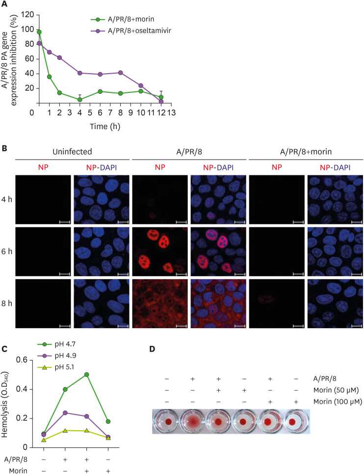
Figure 2. Restriction of entry of influenza virus into host cells by morin hydrate. (A) 250 µM morin hydrate or 10 µM oseltamivir carboxylate was added to A/PR/8-infected A549 cells at the indicated time points. The percentage of viable cells was calculated at 14 h post-infection. Viral RNA was analyzed using RT-PCR. (B) A549 cells were infected with 1×10³ pfu of A/PR/8 and treated with 250 µM morin hydrate. Cells were harvested at 4, 6, and 8 h post-infection, and stained to detect NP under a fluorescence microscope. (C) For the hemolysis inhibition assay, a mixture of 100 µM morin hydrate and 1×10³ pfu of A/PR/8 were added to freshly obtained chicken erythrocytes and incubated at pHs 4.7, 4.9, and 5.1 at 37°C for 30 min. After a brief centrifugation, the released hemoglobin in the supernatants was estimated by measuring the OD at 540 nm. (D) For hemagglutination assay, chicken erythrocytes were mixed with A/PR/8 virus and 50 µM and 100 µM morin hydrate.

Figure 2