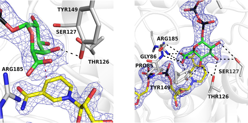
Figure 5.
The crystal structure of BcUGAepi in complex with NAD+ and UDP-galacturonic acid at pH 6.5 and 4 °C shown in two different orientations. The left panel highlights the position of the C4′ in proximity of the nicotinamide (red dashed line) and the hydrogen bond between the sugar O4′ and Tyr-149 (black dashed line). As shown in the right panel, all hydroxyl groups and the carboxylate of the glucuronic acid are engaged in hydrogen-bonding interactions with the protein. The orientations are the same as in Fig. 3 (A and B). The contour level of the weighted 2Fo − Fc density is 1.4σ (subunit A).

