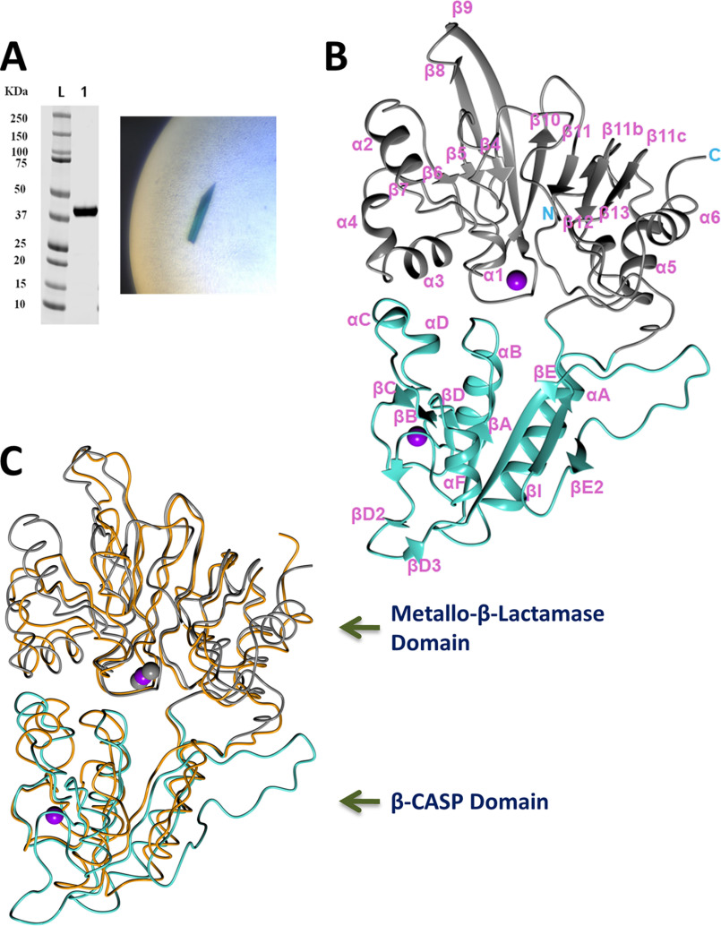
Figure 2.
Crystal structure of Artemis catalytic domain. A, left, purified Artemis 1–368. Right, crystal of crystal form 1 with blue color from Cyanine5 dye. B, overall Artemis structure in ribbon diagram: the metallo-β-lactamase domain is colored gray and the β-CASP domain is colored turquoise. The zinc ions are shown in purple spheres. C, superposition of SNM1B/Apollo (PDB entry 5AHO) in orange for the polypeptide and gray spheres for zinc ions and Artemis colored as in panel B.

