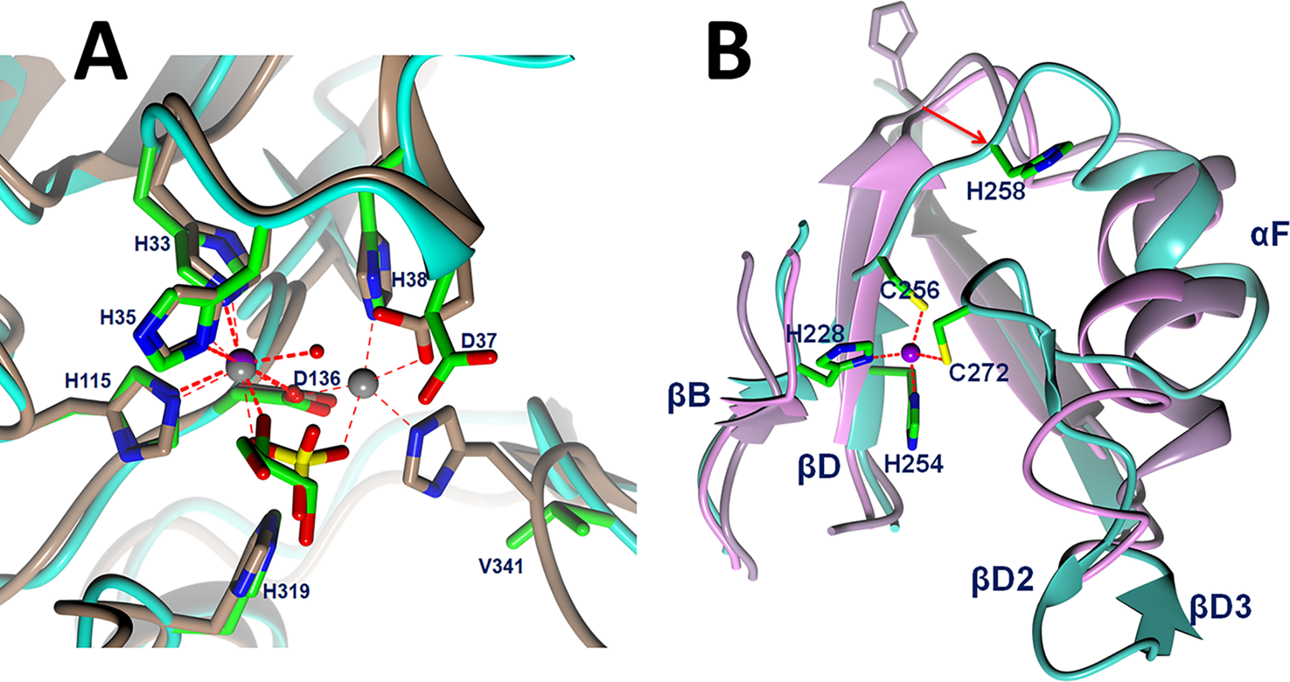
Figure 4.

A, comparison of active site of Artemis with CPSF-73 (PDB entry 2I7T) (8). Zinc ions are shown as a purple sphere for Artemis and gray spheres for CPSF-73. Artemis backbone is shown as a cyan ribbon, and the side chains of conserved residues are shown in green for carbon, blue for nitrogen, and red for oxygen atoms. Water molecules are shown as red spheres. The main chain and side chains of CPSF-73 are shown in brown. A sulfate (yellow sulfur) ion in the CPSF-73 structure and glycerol in Artemis are bound above the catalytic metals in a position likely occupied by the scissile phosphodiester of the substrate. B, novel, structural zinc binding site in Artemis β-CASP domain induced structural differences among Artemis in cyan, SNM1A in pink, and SNM1B/Apollo in lilac. An 8.5-Å movement from SNM1B/Appolo His-234 to Artemis His-258 is elicited as a red arrow.
