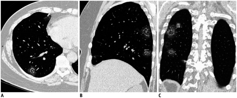
Fig. 2. Chest-CT multi-planar reconstructions of the “double halo sign.”.
Axial (A), sagittal (B), and coronal (C) images show rounded areas of normal parenchyma or rounded ground-glass opacities surrounded by a more or less complete ring of consolidation surrounded in turn by another peripheral ground-glass halo with a final target-like appearance that we defined as “double halo sign.”

