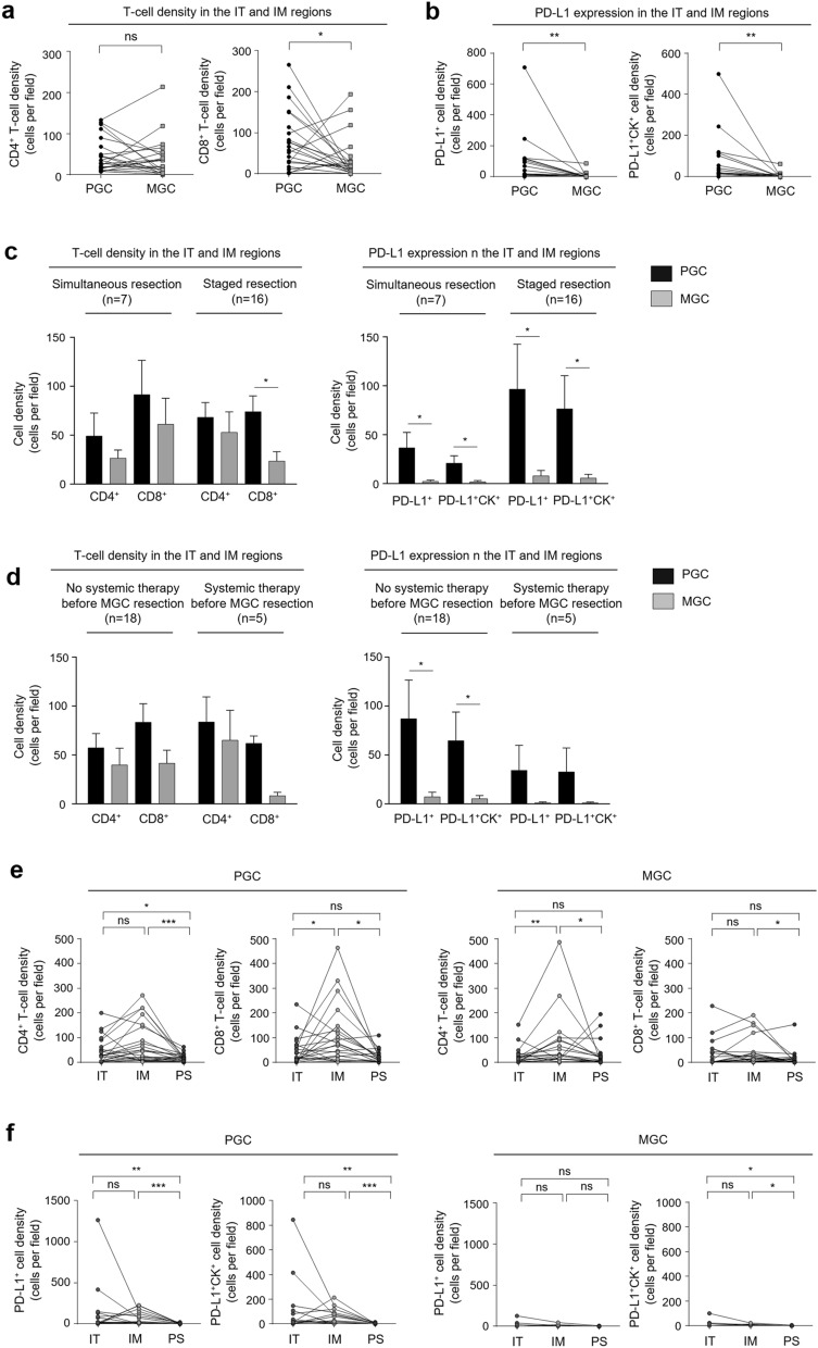
Figure 1.
Tumor-infiltrating T-cell density and PD-L1 expression in PGC and MGC. (a) Comparison of CD4+ and CD8+ T-cell density in the IT and IM regions. (b) Comparison of PD-L1+ and PD-L1+CK+ cell density in the IT and IM regions. (c) T-cell density and PD-L1 expression in the IT and IM regions analyzed according to the duration between gastrectomy and MGC resection and (d) whether patients received systemic therapy before MGC resection. (e) CD4+ and CD8+ T-cell density and (f) PD-L1+ and PD-L1+CK+ cell density according to tumor region. PGC primary gastric cancer, MGC metastatic gastric cancer, IT intratumoral, IM invasive margin, PD-L1 programmed death-ligand 1, CK cytokeratin. Statistical analyses were performed using the Wilcoxon signed rank test (ns not significant; *P < 0.05; **P < 0.01; ***P < 0.001).

