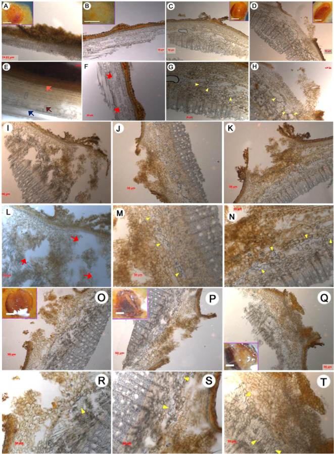
Fig. 2. Anatomy of lenticels during the rooting of apple cuttings.
Each lenticel was observed under a stereomicroscope at 0 h (a, e), 72 h (b–d, f–h), 120 h (i–n), and 168 h (o–t). The treatments are as follows: control (a, e, c, g, j, m, p, s), NPA (b, f, i, l, o, r), and IAA (d, h, k, n, q, t). Scale bars, 50 μm. Inset scale bars, a–d = o–q = 0.5 mm. Arrows: yellow, proliferated founder cells; pink, epidermis; brown, parenchyma cells located in the interfascicular cambium; dark blue, vascular tissues; red, cavities.

