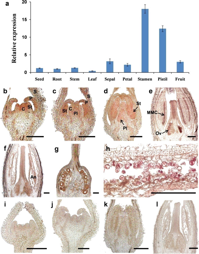
Fig. 2. Expression analyses of SlMYB33 in tomato.
a qRT-PCR analyses of SlMYB33 in different tomato tissues. The various tissues in different developmental stages included in this analysis were as follows: seeds, red ripe fruit stage; roots, stems, and leaves, four true leaf-stage plants; sepals, petals, stamens, and pistils, 5-mm-length flower buds; fruits, 7-DPA (days post anthesis) stage. Values are the mean ± SD of three biological replicates. b–l In situ hybridization of SlMYB33 in tomato flowers at different developmental stages. b–h Longitudinal sections of flower buds hybridized with the antisense probe at stage 4 (b), stage 6 (c), stage 7 (d), stage 9 (e), stage 12 (f), and stage 14 (g, h). i–l Negative controls hybridized with the sense probe (no signals were detected). S sepal, P petal, St stamen, C carpel, Pl placenta, MMC microspore mother cell, Ov ovule, An anther. Bars = 200 μm

