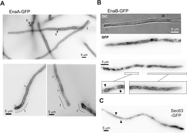
Figure 6.
Subcellular localization of EnaA-GFP and EnaB-GFP. Cells were grown in selective WMM for 16 h at 25 °C and shifted to freshly prepared WMM adjusted to pH 8 with 0.1 M Na2HPO4. Images were taken approximately 1.5 h after the shift. (A) Localization of EnaA along the plasma membrane; in the tip (t), brunches (b), septa (s) and grouped in clusters (white arrowheads). (B) Localization of EnaB in internal membranous organelles. Magnifications correspond to the tip region, where EnaB distributes in a network of strands and tubules, and a more distal region where is localized in ring-shaped structures (black arrowheads). Localization of Sec63-GFP is included as an ER marker. Green fluorescence images are shown in inverted gray contrast. Bars = 5 μm.

