
Figure 2.
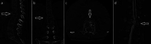
a-c) TC images showing complete collapse of L1with back wall protruding into the spinal canal, in figure c there was bone changes extended to both the pedicles (honey comb pattern). d, MR images showing vertebral body collapse and cord compression.