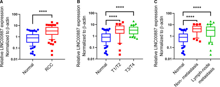
Fig. 1.

LINC00887 expression is frequently increased in RCC tissue. (A) Boxes show LINC00887 expression in 79 pairs of RCC tissues and matched normal tissues. Comparisons were performed with Student’s t‐test. (B) Boxes show LINC00887 expression in 38 T1/T2‐stage RCC tissues, 41 T3/T4‐stage RCC tissues and 79 matched normal tissues. (C) Boxes show LINC00887 expression in 49 lymph node‐metastatic RCC tissues, 30 non‐metastatic RCC tissues and 79 matched normal tissues. Comparisons were performed with one‐way ANOVA followed by Tukey’s post‐hoc test. LINC00887 expression was measured using qRT‐PCR, with β‐actin serving as an internal control. Whiskers: 10–90 percentile. ****P < 0.0001.
