Fig. 3.

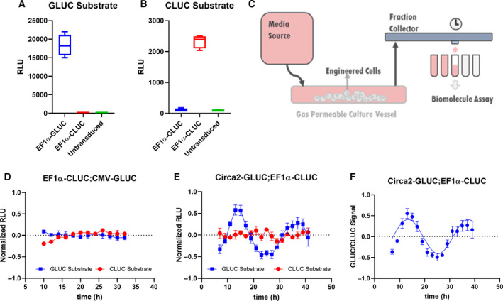
Rhythmic and constitutive secretion of GLUC and CLUC from lymphoblastic leukemic T cells in a suspension cell flow collection system. Media conditioned with EF1α‐GLUC, EF1α‐CLUC, or unmodified (untransduced) Jurkat cells was assayed using (A) GLUC substrate or (B) CLUC substrate separately. (C) Schematic of a continuous flow system in which Jurkat cell secretion was monitored. Jurkat cell secretions were collected from synchronized (D) EF1α‐CLUC; CMV‐GLUC or (E) Circa2‐GLUC; EF1α‐CLUC cells, linear detrended (Period: ~ 21.3 h), and amplitude normalized (SD; N = 6). (F) Circa2‐GLUC signals were normalized to EF1α‐CLUC signals (quotient of amplitude normalized and linear detrended GLUC to CLUC signals) and an undamped 24‐h Sine wave was fitted to the data (Prism, least squares method, degrees of freedom: 14, R squared: 0.7777, sum of squares: 0.4124). Student's t‐test was used to assess significance where relevant.
