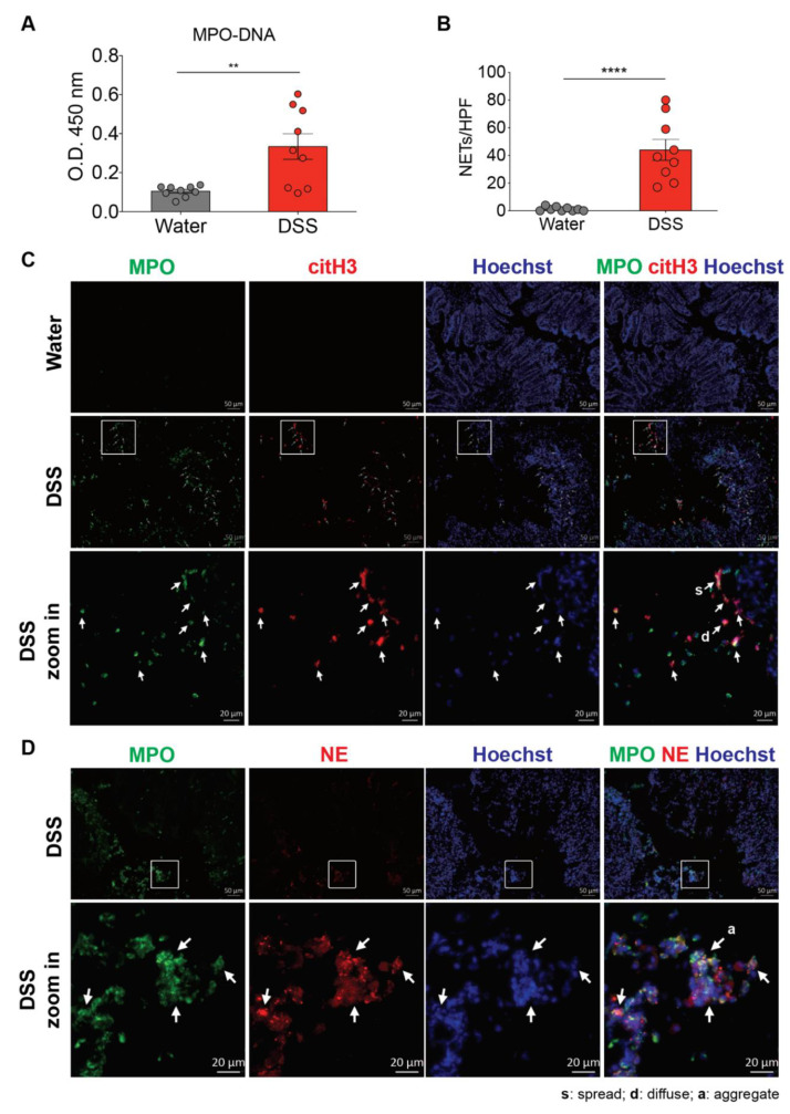
Figure 1.
Neutrophil extracellular traps (NETs) are induced in the colon of dextran sulfate sodium (DSS)-treated mice. (A) NET release was measured in colon homogenates of wild-type C57BL/6 mice 8 d after the consumption of clean water or 2.5% DSS, by using myeloperoxidase (MPO)-DNA ELISA. (The results are pooled data from two separate experiments. n = 9 mice per group.) (B,C) NET formation counts per 200X high-power field (HPF) and representative NET release (white arrows) in the colon of wild-type C57BL/6 mice 8 d after the consumption of clean water or 2.5% DSS, assessed by immunofluorescence microscopy of MPO (green), citrullinated histone H3 (citH3; red), and Hoechst 33342-stained DNA (blue). Bottom row, enlargement of the area outlined in the second-row images. (The results are pooled data from two separate experiments. n = 9 mice per group.) (D) Representative NET release (gray arrows) in the colon of wild-type C57BL/6 mice 8 d after the consumption of 2.5% DSS, assessed by immunofluorescence microscopy of MPO (green), neutrophil elastase (NE; red), and Hoechst 33342-stained DNA (blue). Bottom row, enlargement of the area outlined in the second-row images (n = 9 mice per group). s: NETs in spread form, d: NETs in diffuse form, a: aggregate NET. The data represent the mean ± SEM. Statistical analyses were performed using unpaired, two-tailed t-test. ** p < 0.01, **** p < 0.0001. Scale bars, 50 µm. 20 µm in zoom in pictures.

