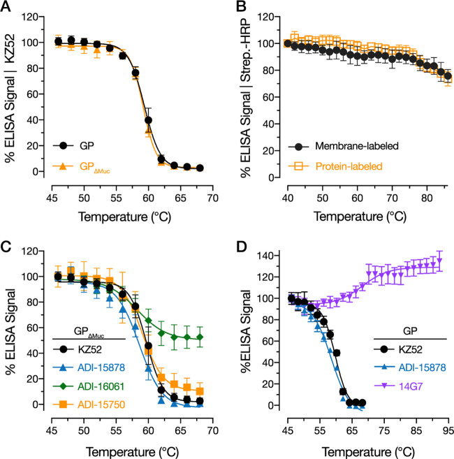
FIG 1.
Thermal denaturation curves for prefusion epitopes in uncleaved EBOV GP. (A) rVSV-GP and rVSV-GPΔMuc were incubated at the indicated temperatures for 10 min, after which the samples were cooled to 4°C and KZ52 binding was assessed by ELISA. Averages ± standard deviations (SD) are shown; n = 9 from 3 independent experiments. Average absorbance range at 450 nm (A450) at lowest temperature tested (46 °C), 2.0 to 2.3. (B) Membrane- and protein-labeled rVSV-GPΔMuc preparations were incubated at the indicated temperatures, and biotin-labeled particles were detected with streptavidin-HRP by ELISA. Averages ± SD are shown; n = 6 from 2 independent experiments. (C) Effect of virus preincubation at the indicated temperatures on binding by MAbs ADI-15878, ADI-16061, ADI-15750, and KZ52. Averages ± SD are shown; n = 15 from 4 independent experiments (except ADI-15878, where n = 12 from 4 independent experiments). Average A450 at lowest temperature tested (46 °C),1.6 to 2.2. (D) Effect of virus preincubation at the indicated temperatures on binding by Muc-specific MAb 14G7. Binding curves for KZ52 and ADI-15878 binding are shown for comparison. Averages ± SD are shown; n = 9 from three independent experiments. Average A450 at lowest temperature tested (46 °C), 0.75 to 1.2.

