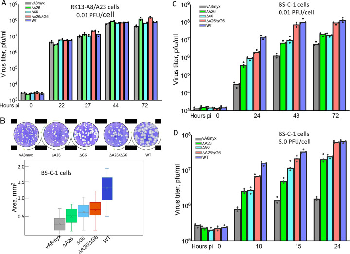
FIG 2.
Plaque formation and replication of vA8myx and A26L and G6R deletion mutants. (A) Replication of viruses in RK13-A8/A23 cells. Cells were infected in duplicate with 0.01 PFU/cell of vA8myx (gray), vA8myxΔA26 (green), vA8myxΔG6 (light blue), vA8myxΔA26ΔG6 (red), or the WT virus (dark blue). At the indicated hours postinfection (pi), virus titers were determined by plaque assay on BS-C-1 cells. (B) Plaque sizes. Crystal violet-stained plates (upper panel) and plaque areas (lower panel) are shown. (C) Virus spread. BS-C-1 cells were infected in triplicate with 0.01 PFU/cell of viruses (data are color coded as described for panel A). At the indicated times, virus titers were determined by plaque assay. (D) Virus yields. BS-C-1 cells were infected in triplicate with 5 PFU/cell of viruses (data are color coded as described for panel A). At the indicated times, virus titers were determined by plaque assay.

