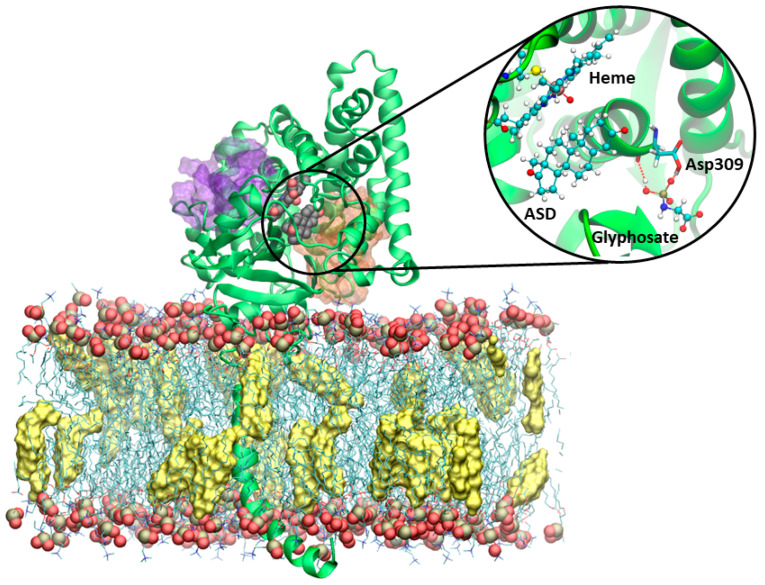
Figure 3.
Representative structure of aromatase embedded in a POPC (1-palmitoyl-2-oleoyl-sn-glycero-3-phosphocholine) membrane, with phosphorous and oxygen atoms shown as tan and red van der Waals (vdw) spheres, and cholesterol (yellow surface) membrane. Sites 1 and 2 are shown as orange and purple transparent surfaces, respectively. The heme and androstenedione (ASD) are displayed in a vdw representation. The protein is shown as green new cartoons. The inset reports a close view of structure of aromatase in complex with glyphosate, as obtained from the most representative cluster of the molecular dynamics simulation trajectory. The heme moiety, ASD, and glyphosate are depicted in balls and sticks. The key catalytic residue Asp309, lining the binding cavity, is shown in licorice and colored by the atom name.

