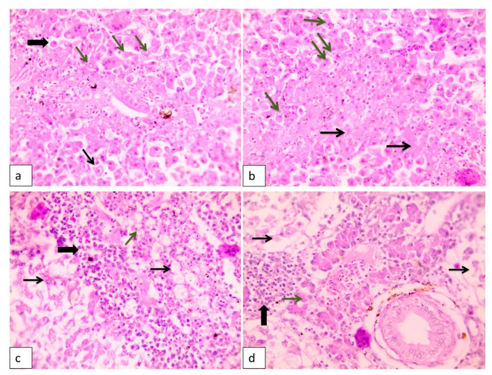
Figure 1.
Histopathological lesions in liver of naturally diseased Nile tilapia showing (a) hepatocellular dissociation (thin arrow) with many necrotic and apoptotic cells (thick arrow). Few rod-shaped bacteria are inside a hepatocyte cytoplasmic vacuole (green arrows). H&E (hematoxylin and eosin), 400×; (b) Widespread hepatocellular necrosis (black arrows) with diffuse, numerous rod-shaped bacteria (green arrows). H&E, 400×; (c) Diffuse, severe necrotizing hepatitis. See widespread, severe hepatocellular lysis (arrows) with few bacteria (green arrow) and widespread infiltrations with numerous macrophages and lymphocytes (thick arrow). H&E, 400×; (d) Hepatopancreatitis with edema widely separated lytic hepatocytes (arrows) and lysis of hepatopancreas (green arrow) and focal aggregations of inflammatory cells (thick arrow).

