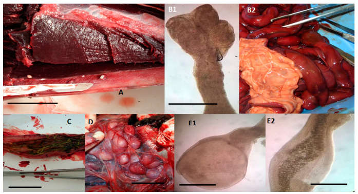
Figure 3.
Cestodes and Acanthocephalans identified in dolphins (15 Stenella coeruleoalba, 10 Tursiops truncatus, and a Grampus griseus), stranded along the Tuscan coastline (central Italy) of the “Pelagos Sanctuary” in the period between February 2013 and July 2015. (A) Phyllobothrium delphini located in the subcutaneous adipose tissue of the perigenital region of a striped dolphin (S. coeruleoalba), macroscopic view. Merocercoid larvae appear as white oval cystic formations with a diameter of 5–10 mm and containing an invaginated scolex showing four grooves (bothria) and a short neck, scale bar 2 cm. (B1,B2) Tetrabothrium forsteri adults in the intestine of a striped dolphin (S. coeruleoalba), microscopical view of the scolex showing four bothria measuring 0.3–0.69 mm in length and 0.25–0.6 mm in width (B1, scale bar 1 mm), and macroscopic view of the strobila whose length may range from a few millimetres to two metres, while the proglottids are wider than long (B2). (C) Strobilocephalus triangularis adults in the intestine of a striped dolphin (S. coeruleoalba). The size of the strobila varies from a few millimetres to two meters, while the scolexes are 5–6 mm wide and 4–6 mm long with four muscular bothria (scale bar 2.5 cm). (D) Monorygma grimaldii merocercoids in the subserosa of the abdominal cavity of an infected striped dolphin (S. coeruleoalba). Merocercoids appear as white cystic formations with a diameter of 10–20 mm, each containing an invaginated scolex showing four bothria and a very long neck, scale bar 1.5 cm. (E1,E2) Bolbosoma vasculosum female adult specimen about 0.435 mm wide and 0.85 mm long, found in the intestine of a bottlenose dolphin (T. truncatus) showing the bulbar anterior end of the body (E1, scale bar 250 µm) and developed eggs (E2, scale bar 250 µm).

