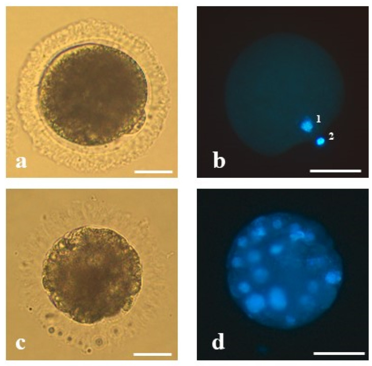
Figure 1.
The representative oocyte and morula stage embryo of the domestic cat analyzed in the present study: (a) mature oocyte of proper morphology at the second metaphase stage with extruded first polar body, (b) mature oocyte stained with Hoechst 33,342—(1) visible chromatin of the oocyte and (2) the first polar body, (c) an embryo at the morula stage on 5 dpi derived from an oocyte matured in vitro, vitrified, fertilized by intracytoplasmic sperm injection and (d) stained with Hoechst 33,342. Bars represent 50 µm.

