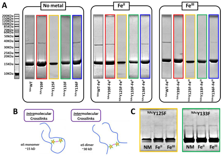
Figure 5.
SDS-PAGE analysis of PICUP Samples. (A) Samples from PICUP reactions with NAcαSyn variants supplemented with no-metal, FeII, or FeIII separated on 10% SDS-PAGE gels and stained with Coomassie blue R-250. Ladder is located to the left of the gels. (B) Schematic representation of intramolecular and intermolecular dityrosine crosslinks. (C) Concentrated samples of PICUP reactions performed with NAcY125F (yellow) and NAcY133F (green) separated on 10% SDS-PAGE gels stained with Coomassie blue R-250.

